Cyril: Large Field of View Widefield microscope
Summary
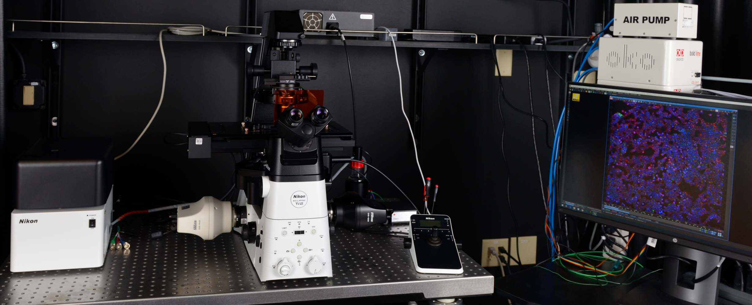
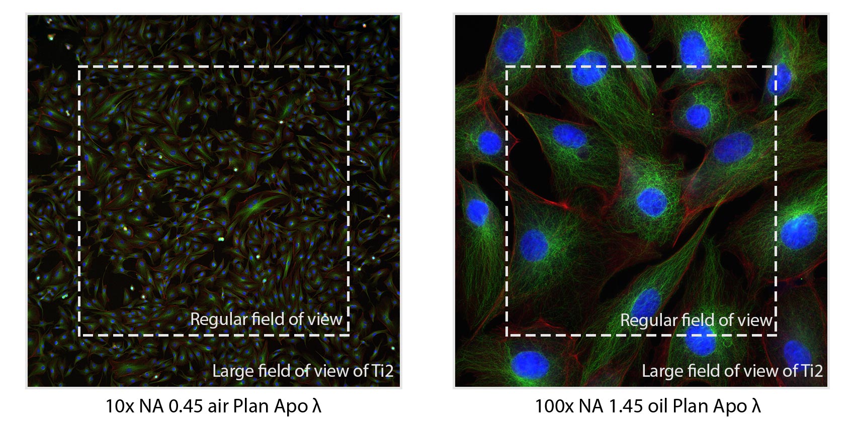
Cyril is a widefield microscope with a large field of view and is equipped for 4 imaging modalities: brightfield, Differential Interference Contrast (DIC), color (RGB), and widefield fluorescence. The biggest advantage of this system is the large field of view; every imaging mode benefits from this. This large field of view shows even multiple cells with subcellular details at 100x magnification! Cyril is excellently suited for fixed and live cells. Complete automation in combination with perfect focus system make this an ideal system to acquire entire slides and 96 well plates in an unattended fashion. This could even be done as time lapse with a duration of hours or even overnight and longer.
Image modalities
- Brightfield
- DIC imaging
- Color imaging
- Widefield fluorescence imaging
Applications
-
Histology. The color imaging mode is great for fixed tissue slices stained with e.g. hematoxylin and eosin (HE), Periodic acid-Schiff reaction (PAS), Masson's trichrome, Azan stain, and Giemsa. NIS elements software allows easy white balance calibration. We can create an automated acquisition pipeline (JOBS) to acquire mulitple slides in one run.
-
Immunofluorescence. There are 6 different LED colors with matching filters available in the widefield fluorescence imaging mode.
-
Fluorescence live cell imaging. In the widefield fluorescence mode there are 6 different LED excitation colors available which are matched with appropriate filters. The stage top incubation chamber regulates temperature, CO2 and humidity.
-
Organoid imaging. The large field of view is ideal to acquire an entire organoid in one frame. In addition, long working distance objectives are available to image through the entire organoid.
-
DIC and brightfield. DIC and brightfield can be used as standalone image modes or can be combined with fluorescence or color imaging. The stage top incubation chamber is fully DIC compatible.
Technical description
|
|
Specifications |
|
Microscope body |
Eclipse Ti2-E |
|
Stage |
Automated |
|
Perfect Focus system (PFS) |
Yes |
|
Incubator |
Okolab Stage top (25–50⁰C, CO2, humidity including objective heater and temperature sensor) |
|
FOV |
25mm (D) |
|
Objectives |
Correction, Magnification, Numerical Aperture, Working Distance |
|
Plan Apo ʎ 4x NA 0.20 air Plan Apo ʎ 10x NA 0.45 air WD 4000µm Plan Apo ʎ 20x NA 0.75 air WD 1000µm Plan Apo ʎ 40x NA 0.95 air Plan Fluor 40x NA 1.30 oil WD 240µm Plan Apo ʎ 60x NA 1.40 oil WD 130µm Plan Apo ʎ 100x NA 1.45 oil WD 130µm 10x, 20x, 40x, 60x and 100x magnifications are compatible with DIC and Brightfield |
|
|
Software |
NIS-Elements software (version 5.42.03 High content analysis package) |
|
RGB Imaging |
Diascopic LED |
|
Fluorescence Imaging |
LED: 395/25nm, 440/20nm, 470/24nm, 510/25nm, 550/15nm, 640/30nm (Lumencor SpectraX) |
|
Detectors |
DS-Ri2 Camera/DS-Qi2 CMOS 16.25megapixels (not interpolated) 4908 x 3264 pixel (full-pixel) images at 6 fps, or 1636 x 1088 pixel (3 x 3 pixel averaging) images at 45 fps Imaging format 36mm x23.9mm/6.91 x 4.92mm |
|
Emission filters |
DAPI 460/25nm FITC 535/40nm Cy5 635/53nm Cy7 700/75nm Triple emission filer (CFP/YFP/mCherry, Semrock) Quad emission filter (LED-DA/FI/TR/Cy5-B, Semrock) 432/36nm, 415/31nm, 596/31nm, 730/139nm |
Location
Leichtag building, Room 470