JOBS: fully automated acquisition pipeline
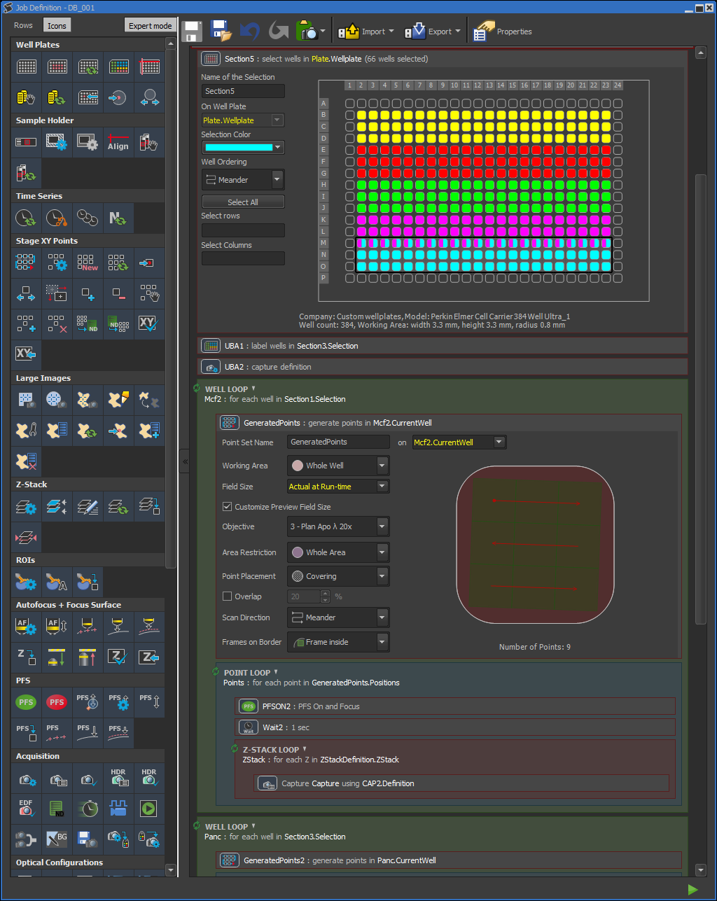
We can help you further streamline your microscopy research by creating an automated acquisition pipeline (JOBS) tailored to your project. We can do this on any of our microscopes. For Krieger, it is the only method to acquire data. JOBS is a module of the NIS-Elements software. It is best described as a method to graphically program an automated acquisition, virtually any kind of automated acquisition is possible (see figure). This method is ideal to perform high content screenings, but it can be applied to almost any type of microscopy projects. Below you see a few examples of workflows that we can create.
High content live cell imaging JOBS
- User mounts a 384 well plate in the stage top incubator.
- User selects the wells to be imaged. Within the same multi-well plate, you can image multiple groups of wells with different acquisition settings. These different groups are color coded in the example figure.
- User selects how many positions in each well will be acquired.
- The NIS-Elements software acquirers at every position, for example a Z-stack for a confocal image for DAPI, Alexa488, Alexa560 and Alexa640.
- The NIS-Elements software loops over every position every 30 minutes for 16 hours.
- This results in a hyper stack which contains a Z stack for each channel (DAPI, Alexa488, Alexa560 and Alexa640) at every timepoint at every position.
Histology JOBS
- User loads 4 slides.
- The NIS-Elements software acquirers overview of 4 slides with low magnification
- User selects region of interests to image
- The NIS-Elements software generates point within region of interest
- The NIS-Elements software acquires at all points color images within the region of interests multiple Z-planes using high magnification.
- The NIS-Elements software stiches the focused images together.
- Result stitched high-resolution color image per region of interest.
Single molecule tracking JOBS
- User mounts a multi chamber slide in the stage top incubator chamber.
- User positions the cells.
- The NIS-Elements software acquirers one frame in the green channel TIRF mode (membrane marker).
- The NIS-Elements software acquirers a time-series in the red channel TIRF mode (single molecules of interest), continuously imaging with 30 ms per frame for 5 min.
- The NIS-Elements software acquirers one frame in green TIRF mode (membrane marker).
- The NIS-Elements software combines all images in one hyperstack. The green channel can be used as outline for the membrane and individual molecules can be tracked in the red channel.