ACTRI AX R: Confocal
 Summary
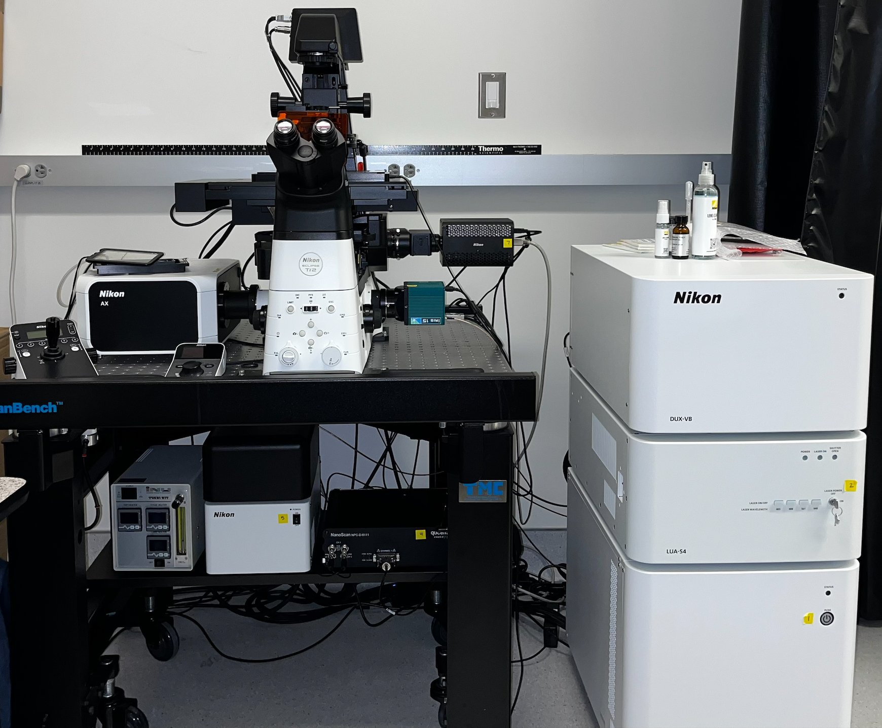
Nikon’s newest point scanning confocal offering an industry leading 25mm field of view, hybrid spectral and filter-based detection, up to 8K resolution galvo scanning and 2K high speed resonant scanning with 405/488/561/640 excitation. Also offers incubation, DIC imaging, fast Z-section capabilities, and automatic water dispenser for high-throughput, high-resolution experiments.
Imaging modalities
- Laser scanning confocal – Galvano scanner and Resonant scanner
- Spectral imaging
- Bright field and DIC
Applications
- Immunofluorescence samples. Imaging entire slides is easy and straightforward using tiling and stitching functions in the NIS-Elements software. You can control the orientation of the Galvo scanner and thereby control the orientation of your sample within the image.
- Live cell imaging. Tokai Hit stage top incubation chamber regulates temperature (25 – 50 ⁰C), CO2 and humidity. The resonant scanner is ideal to image fast moving processes with bright fluorophores.
- FRAP and stimulation experiments. You can image with the resonant scanner and use the Galvo scanner to simultaneously stimulate, activate or convert with the 405 nm laser simultaneous. Or you can bleach, activate or stimulate with the 405 nm, 488 nm, 561 nm or 640 nm laser and image sequentially using only the Galvo scanner. The stimulation function is integrated in the NIS elements software. Cells can be immediately analyzed after or even during the acquisition with the time measurement tool.
- Spectral imaging. The spectral range is 400 – 750 nm with a bandwidth of 5, 10, 20, 40nm with a range of 9-70 channels depending on bandwidth.
- Filter / ratiometric FRET. Filter FRET using blue, green, red and far red fluorescent dyes or fluorescent proteins can be performed. You could measure a FRET change of a cytosolic FRET sensor and acquire multiple cells in one field of view at low magnification or you could measure at high magnification thereby observe a FRET change at a specific subcellular localization.
- Spectral FRET. Similar as filter / ratiometric FRET, but with more freedom of different colors of fluorescent probes, since you can measure the entire spectrum from 400 – 750 nm with a bandwidth of 5, 10, 20, 40nm
- Image ‘exotic’ and large Stokes shift fluorophores. The tunable emission width in combination with 405, 488, 561 or 640 nm lasers provide many acquisition options for a wide variety of fluorophores.
Technical description
|
|
Specifications |
|
Microscope body |
Eclipse Ti2-E |
|
Stage |
Automated |
|
Piezo Drive |
Queensgate 600µm |
|
Perfect focus system (PFS) |
Yes |
|
Incubator |
Tokai Hit Stage top (25 – 50 ⁰C, CO 2, and humidity) |
|
Objectives |
Correction, Magnification, Numerical Aperture, Working Distance |
|
Plan Apo ʎ 4x N.A. 0.2 air, WD 20000 µm Plan Apo ʎ 10x NA 0.45 air, WD 4000 µm Plan Apo ʎ 20x NA 0.75 air, WD1000 µm Plan Apo ʎ 40x NA 1.15 water, WD 590-610 µm Plan Apo ʎ 60x NA 1.40 oil, WD 130µm |
|
|
Software |
NIS Elements software (version 5.42.02: High content analysis package) |
|
Lasers |
405 nm, 488 nm, 561 nm, and 640 nm (LUA-S4) |
|
Scanning modalities |
Galvano- up to 8k frame size Resonant- up to 4k frame size |
|
Detectors |
DUX-VB 418-475 nm multi-alkali PMT 400-750nm GaAsP variable range (typically 499-543 nm, 571-625 nm) 655-850 nm GaAsP IRIS 15 15 Megapixel Camera 4.25 µm x 4.25 µm Pixel Area Large 25 mm Diagonal Field of View |
|
Filters |
DAPI filter- 447/57 CFP filter- 482/35 Cy5 filter -640 (Short Pass. For reflecting 640 emission to fixed detector.) Cy7 filter- 730 (Short Pass. For reflecting 730 emission to fixed detector.) |
|
Dichroic mirrors |
405/488/561/640 |
|
Beam splitter |
80/20 |
|
Spectrum scanning |
400 – 750 nm bandwidth of 5, 10, 20, 40nm 9-70 channels 32PMT detectors |
Location
ACTRI L2-W420